The equine hock is a powerful, complex structure that plays a vital role in hind-limb movement, propulsion, and athletic performance. Made up of multiple joints, tendons, ligaments and synovial bursae, it is an area frequently involved in lameness and performance-related issues. This guide from Avonvale Equine Practice explains the anatomy of the hock, how it functions, and the conditions we commonly diagnose in this region.
The Joints of the Hock
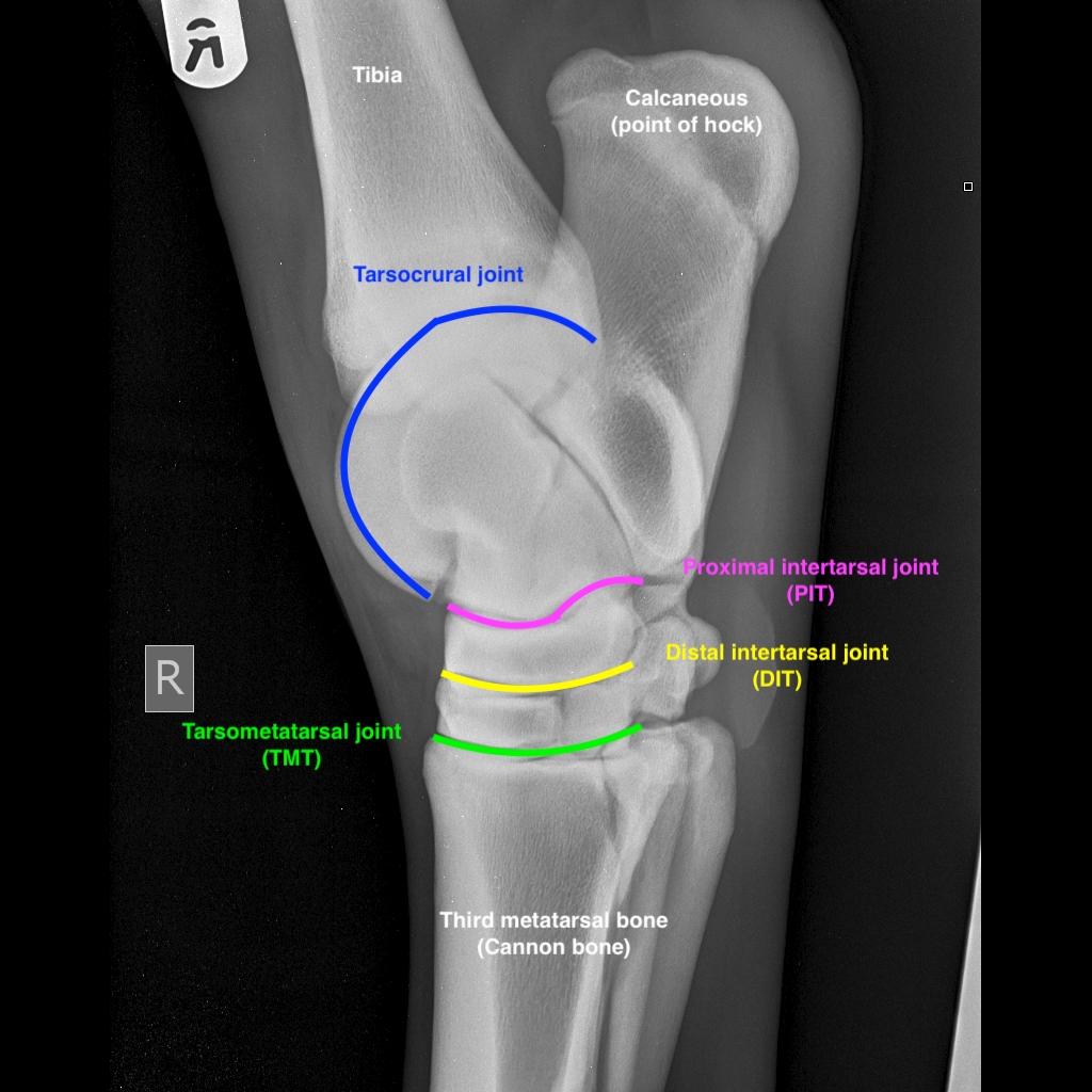
The hock contains four joints, but only one is responsible for significant movement:
- Tarsocrural joint — the main high-motion “hinge” joint that allows flexion of the hock.
- Proximal intertarsal joint (PIT)
- Distal intertarsal joint (DIT)
- Tarsometatarsal joint (TMT)
The PIT, DIT and TMT joints are low-motion joints, meaning they contribute little to flexion but are common sites of arthritis. The DIT and TMT joints are the most frequent locations for degenerative joint disease and are often treated with corticosteroid medication.
Young horses may develop Osteochondrosis dissecans (OCD) in the tarsocrural joint, typically presenting as bone fragments.
Diagnosis of joint pathology usually involves:
- Joint blocks (local anaesthetic injections)
- X-rays to assess bone and joint surfaces
Tendons of the Hock
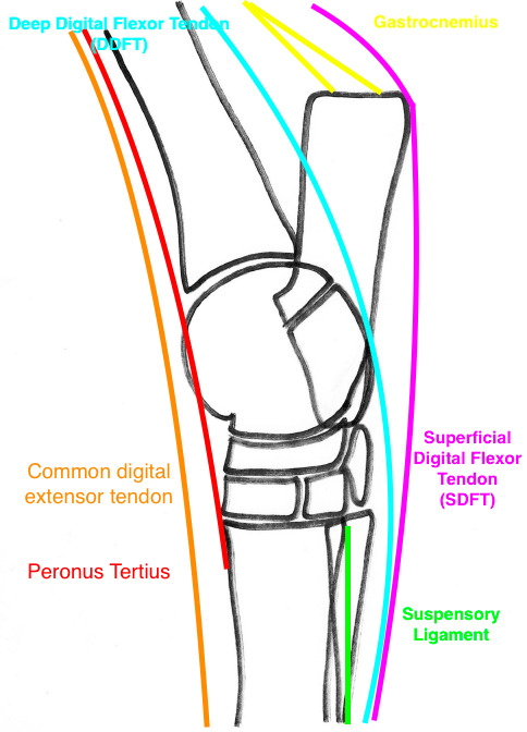
Several important tendons run down the back and front of the hock, contributing to flexion and extension of the limb.
Flexor Tendons (Back of the Hock)
- Deep Digital Flexor Tendon (DDFT) — runs from inside the point of the hock down to the pedal bone.
- Gastrocnemius tendon — part of the Achilles tendon, attaching at the point of the hock.
- Superficial Digital Flexor Tendon (SDFT) — runs over the point of the hock and down the limb.
Injuries to these tendons at hock level are uncommon, but the SDFT can occasionally dislocate if its attachment at the point of the hock is damaged.
The bursae around the point of the hock can swell or become infected:
- Capped hock — swelling of the superficial bursa
- Deep bursitis — often associated with wounds over the point of the hock
Extensor Tendons (Front of the Hock)
- Common Digital Extensor Tendon — runs down the front of the limb; horses can recover well even if this tendon is cut.
- Peroneus tertius — part of the hind-limb stay apparatus, linking the stifle and hock.
- If ruptured, the horse can extend the hock while the stifle remains flexed — a key diagnostic sign.
Ligaments of the Hock
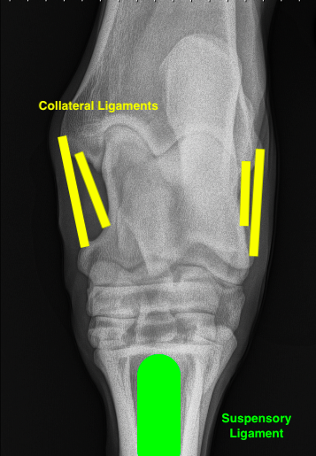
Ligaments stabilise the hock and support the limb during movement.
- Collateral ligaments — located on either side of the tarsocrural joint, preventing twisting.
- Suspensory ligament — originates just below the TMT joint and supports the fetlock and pastern.
Collateral ligament injuries are rare but may occur following a kick and often require prolonged rest.
Proximal suspensory ligament desmitis is a common condition in sports horses, diagnosed via nerve blocks and ultrasound. It may occur alongside low-grade hock arthritis.
Diagnosing Hock Problems
If your horse is lame or has sustained a hock injury, a thorough assessment is essential. At Avonvale Equine Practice, we use:
- X-rays — to evaluate bones and joint surfaces
- Ultrasound — to assess tendons and ligaments
- MRI — occasionally used for complex or unclear cases
- Diagnostic arthroscopy — keyhole surgery to examine and treat the tarsocrural joint
Early diagnosis allows for targeted treatment and greatly improves long-term soundness.
The Importance of Early Intervention
The hock is placed under significant stress during movement, especially in performance horses. Conditions affecting the hock are not uncommon, but with prompt diagnosis and appropriate treatment, most horses return to comfortable, successful work.
If you have concerns about your horse’s mobility, performance or comfort, the team at Avonvale Equine Practice is here to help.
Register Your Horse/s At Avonvale Equine
If you are looking for an equine vet for your horse/s that delivers high quality routine and 24/7 in-house emergency care, why not get in touch with us? We look after all types of horses, ponies, donkeys and mules within our coverage area, which includes large parts of Oxfordshire, Warwickshire, Northamptonshire, Birmingham, The Midlands as well as areas of Worcestershire, The Cotswolds, and Gloucestershire.
Our experienced and skilled equine vets are passionate about helping both commercial and individual owners look after their equines excellent equine veterinary care, which includes the identification and treatment of sarcoids.
To register your horse with us, complete our new client registration form online or call our practice on 01295 670 501.





.jpg)