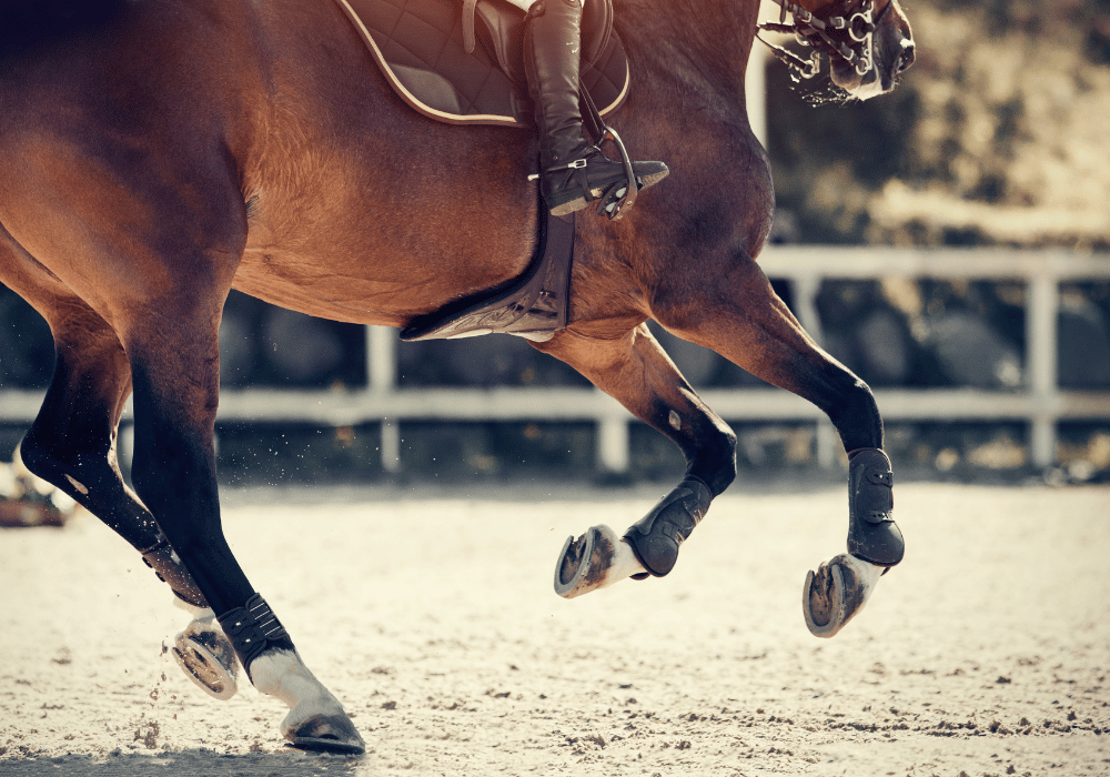
Equine distal limb anatomy is fascinating, but it can also be tricky to grasp. You will probably have heard your vet talk about the various bones, tendons and ligaments, perhaps using their scientific names. This can be confusing to those not familiar with anatomy, so we have created this guide to help horse owners understand the equine distal limb and demystify some of the terminology.
What is the Distal Limb?
The distal limb refers to the horse’s lower leg, below the carpus (knee) or hock.
Around 50 million years ago, Eohippus - an ancestor of the modern horse - walked on five toes. Over time, two toes disappeared completely, whilst the other two evolved into what are now the splint bones.
The modern horse (Equus) is a perissodactyl unguligrade, meaning it walks on hooves and on one digit. This is akin to humans walking on one toe, and understanding this can help you to better understand the horse’s lower leg anatomy.
Thoracic vs Pelvic
The horse’s front and hind legs differ in terms of their anatomy and how they help the horse move and bear weight. The front legs are known as the thoracic limbs, and the hind legs are known as the pelvic limbs.
Muscles in the Horse’s Legs
There are no muscles in the horse’s lower leg. Muscles in the upper leg (the parent muscles) operate long tendons, which extend down the distal limb. These muscles and tendons work together to move the lower leg.
The horse has evolved to not have any muscles in the distal limb to lighten the lower leg and allow the horse to move faster. This helps the horse to run away from predators in the wild. However, the long tendons in the distal limb are vulnerable to injury as a result.
Joints in the Equine Distal Limb
The joints in the horse’s lower leg differ slightly between the thoracic and pelvic limbs. In the thoracic limb, the horse has a knee, whereas in the pelvic limb, this is replaced by the hock.
Hock (Tarsal Joint)
This distinctive joint is characterised by the protrusion of the calcaneus (also known as the point of the hock). The hock has four points of articulation: the tibiotarsal joint, the proximal and distal intertarsal joints and the tarsometatarsal joint.
The tibiotarsal joint, which is the highest joint in the hock, is responsible for the vast majority of the movement. However, that is not to say that the other joints are unimportant. They help absorb impacts and can be prone to injury just like any other joint.
Knee (carpus)
In terms of the bone structure, the carpus is actually more akin to a human wrist joint than a human knee. There is no patella (kneecap), but instead there are two rows of small carpal bones, which fit together almost like a jigsaw puzzle.
However, unlike the human wrist, the horse’s carpus does not have a full range of motion. Instead, it has evolved to be stable enough to support the horse’s weight when standing, whilst being flexible enough to allow the horse to travel at speed. Furthermore, the carpus has to support the horse’s entire body weight on a single limb as the horse canters or gallops.
The carpus is made up of three smaller joints: the antebrachiocarpal joint, the middle carpal joint and the carpometacarpal joint.
Note: The horse does have a patella, which can be found in the stifle joint.
Fetlock (metacarpophalangeal), Pastern (proximal interphalangeal) and Coffin (distal interphalangeal) Joints
This series of joints allow for the articulation of the cannon bone, pasterns and coffin / pedal bone. The flexion and extension of these joints allows the horse to move and absorb concussive force as the hoof hits the ground, and a degree of lateral movement allows the horse’s leg to cope with uneven or rutted ground.
These joints are articulated using the deep digital flexor tendon (DDFT) and the common digital extensor tendons, which originate from the parent muscles much higher up in the horse’s leg.
The Major Tendons in the Distal Limb
Tendons connect muscles to bones. They transmit energy and allow the muscles to move the connected bones. There are two main groups of tendons in the distal limb: the flexor tendons, which flex the limbs and allow them to bend, and the extensor tendons, which help to straighten the legs.
These tendons can be prone to injury - either through knocks or overreach, or through long-term wear, tear and overuse. The tendon sheaths, which protect the tendons as they run across the horse’s joints, can also be vulnerable to damage or infection.
Superficial Digital Flexor Tendon (SDFT)
Injuries to the SDFT are common due to its situation at the back of the leg, as well as the heavy demands placed on the tendon. The SDFT emerges from the superficial digital flexor muscle, extending past the knee or the hock and ending at the short pastern (middle phalanx). The SDFT facilitates flexion of the fetlock and pastern joints.
Deep Digital Flexor Tendon (DDFT)
The DDFT lies beneath the SDFT in the distal limb and also acts to flex the lower leg . Injuries to the DDFT are less common than to the SDFT. The DDFT is particularly vulnerable to external injury in the foreleg where it runs behind the short pastern to attach to the pedal bone. Here, it is more exposed to overreach strikes coming from the horse’s back hooves.
Digital Extensor Tendons
The digital extensor tendons effectively do the opposite to the flexor tendons. They run down the front of the leg and help to extend the horse’s lower leg.
In the thoracic limb, the common and lateral extensor tendons extend the carpus and the leg. In the pelvic limb, the long and lateral extensor tendons extend the digit and flex the hock to bring the hind leg forward.
Ligaments
Ligaments connect bones to other bones. They help to support and stabilise the joints and can limit range of motion to prevent injury. There are many short ligaments throughout the horse’s leg, but this guide will focus on the suspensory ligament and the accessory (check) ligaments.
The Suspensory Ligament
The suspensory ligament is present in the thoracic and pelvic limbs. Its main function is to prevent overextension of the fetlock. The suspensory ligament connects the cannon bone and carpal / tarsal bones to the sesamoid bones.
Accessory (Check) Ligaments
The check ligament most commonly referred to is the inferior check ligament of the forelimb which originates at the back of the knee and travels down the lower leg between the DDFT and suspensory ligament before combining with the DDFT halfway down the cannon. Check ligaments can be susceptible to strain, particularly in older horses.
Major Bones in the Distal Limb
There are several bones in the equine distal limb, including the main cannon bone, splint bones, several small carpal and tarsal bones, sesamoid bones and the pastern and pedal bones.
Cannon Bone
The Cannon Bone is flanked by two splint bones, which usually fuse to the cannon bone as the horse grows.
The cannon bone is properly known as the third metacarpal (in the thoracic limb) or the third metatarsal (in the pelvic limb), whilst the splint bones are the second and fourth metacarpals / metatarsals. What would have been the first and fifth metatarsals and metacarpals have disappeared completely through evolution. The cannon bone connects the hock or knee to the fetlock.
Pastern Bones (Proximal and Middle Phalanxes)
The long (proximal phalanx) and short (middle phalanx) sit between the cannon bone and the pedal bone. After the hoof, the pastern bones are the first structures to absorb concussion as the horse moves.
Coffin Bone (Distal Phalanx)
The coffin / pedal bone (distal phalanx) is the bone inside the horse’s hoof. It is also the bone that rotates and destabilises in cases of laminitis. The distal phalanx is a bit like the bone in the tip of your finger or toe, and this comparison makes it easier to imagine why it’s so important to look after your horse’s hooves! A huge amount of weight is being supported by a relatively small bone, which is also the first to come into contact with the ground, absorbing a great deal of force with every stride.
Avonvale Equine Vets | Equine Orthopaedic Vets
Hopefully, this guide shines a light on how your horse’s legs work and illustrates just how complex they are! Lameness can be very subtle, and the cause is often far from obvious. With so many bones, joints, tendons and ligaments in the distal limb - identifying and treating orthopaedic issues can be complicated.
At Avonvale Equine Vet Practice, our equine orthopaedic experts are fully qualified and equipped to carry out lameness workups, diagnostics and treatments. Our full surgical facilities allow us to carry out a range of procedures, and our experienced team of equine veterinary nurses provide excellent care to our inpatients.
Our equine vet practice covers a wide area surrounding our clinic near Banbury, including Warwickshire, Worcestershire, the Cotswolds and parts of Northamptonshire. Our free weekly zone visits are ideal for routine and non-emergency appointments. Should you require an emergency equine vet, our emergency call-outs are always covered by one of our own vets, 24 hours a day. Register your horse or pony with our equine vet practice today.


