MRI is an imaging technique that measures tissue responses to magnetic fields. These responses can be mapped to produce detailed 3-dimensional anatomical images. This imaging modality has become particularly useful in horses, especially regarding feet, the soft tissue structures within which cannot be seen via radiography or ultrasonography. This case study explains how MRI was used to identify the cause of intermittent lameness in our client's horse.
How is MRI a Useful Diagnostic Tool?
MRI can be valuable in the diagnosis of distal limb issues. Lameness can be complex and difficult to diagnose, and x-rays do not show us everything. In the case of this 10 year-old Irish Sport Horse, an MRI scan proved crucial in getting to the bottom of his intermittent right-forelimb lameness.
The horse was intermittently lame on the right rein only. Some days, the horse could be up to 4/10 lame, and on others, no lameness was visible at all. The owner and rider had assumed initially that it was a bridle lameness. However, they called us and our vet, Emily, carried out a lameness investigation.
An abaxial sesamoid nerve block (which blocks out the foot and pastern) successfully abolished the lameness. X-rays were taken of the horse's front feet, but were found to be unremarkable. Due to the horse's flat foot conformation, Emily suspected that a soft tissue injury within the foot was likely to be the cause and offered the choice of an MRI, or a trial medication of either the coffin joint or the navicular bursa with remedial farriery. The owner elected for the MRI, and the horse was referred to the RVC for an MRI of both front feet.
Interesting Results from the MRI
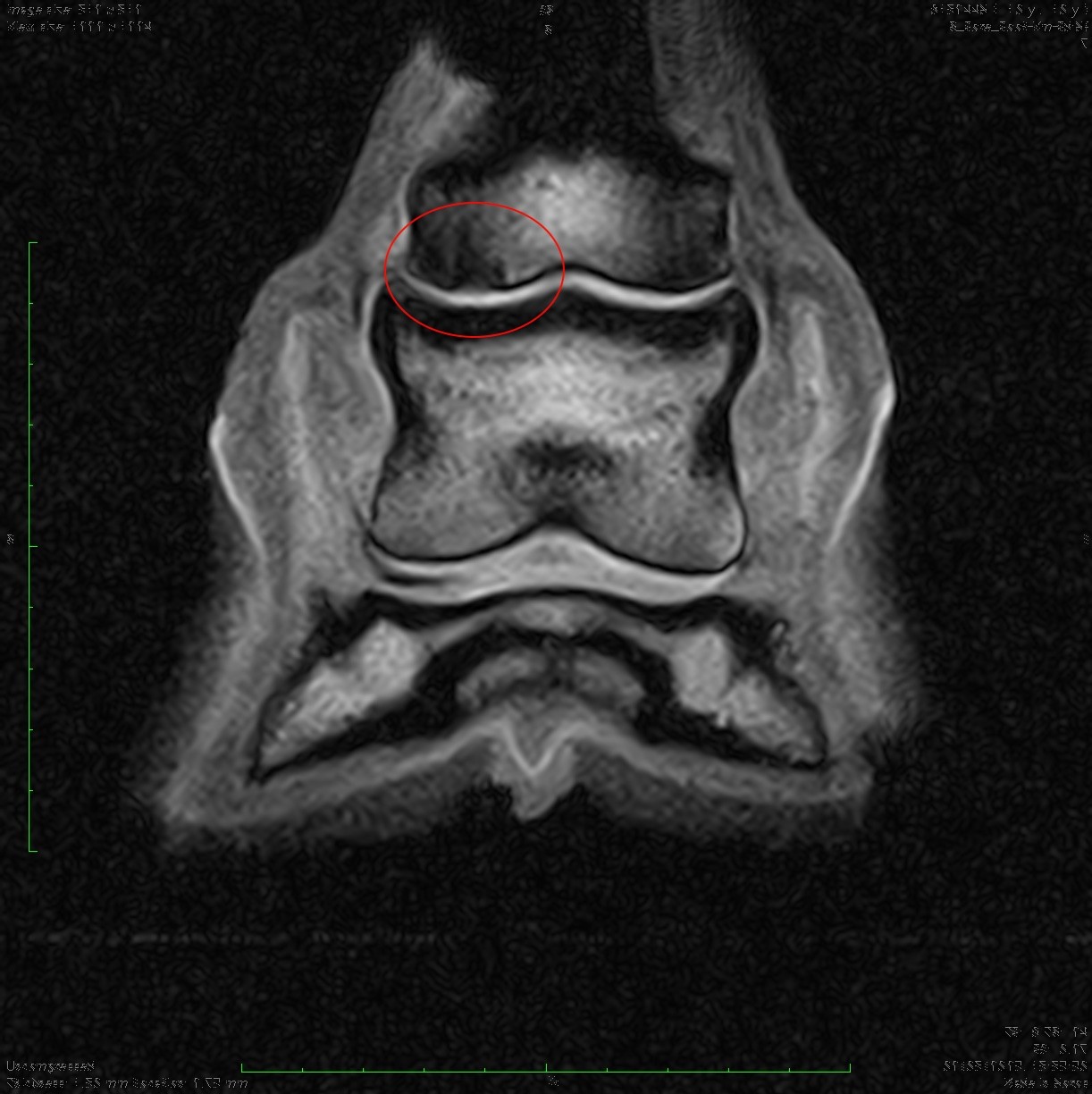
Interestingly, the pastern, rather than the foot, was the area of interest. A chronic defect in the subchondral bone of the lateral condyle of the long pastern was identified, with some minor osteoarthritis of the pastern joint. On its own, minor osteoarthritis of the pastern joint is not often significant, but the underlying damage to the long pastern bone certainly was. Its location is also likely why this horse only appeared lame when on the right rein.
The MRI was an important factor for this horse, as neither a trial medication of the coffin joint nor the navicular bursa would have treated this lesion. Over time, bone lesions like this can deteriorate and predispose the bone to fracturing. The horse is now undergoing treatment and rehabilitation for this condition, thanks to the results gained by the MRI.
Lameness Investigations at Avonvale Equine Vet Practice
We are an independent equine vet practice based in Ratley, near Banbury. Our experienced and qualified vets are well-equipped with the latest diagnostic equipment and we can carry out lameness exams and poor performance investigations at your yard or our clinic. However, we can also refer your horse onto equine hospitals for more advanced or specialist investigations and procedures. Our practice caters for all equines, including leisure horses, companion ponies and competition horses. Register your horse, pony, donkey or mule with us today.